39 dissecting microscope diagram with labels
Label Microscope Diagram - EnchantedLearning.com Using the terms listed below, label the microscope diagram. arm - this attaches the eyepiece and body tube to the base. base - this supports the microscope. body tube - the tube that supports the eyepiece. coarse focus adjustment - a knob that makes large adjustments to the focus. diaphragm - an adjustable opening under the stage, allowing ... Microscope Parts and Functions With Labeled Diagram and Functions How ... Body tube (Head): The body tube connects the eyepiece to the objective lenses. Arm: The arm connects the body tube to the base of the microscope. Coarse adjustment: Brings the specimen into general focus. Fine adjustment: Fine tunes the focus and increases the detail of the specimen.
essayhelpp.com › biology-hBIOLOGY H - Essay Help Mar 07, 2022 · Provide names to the labels of parts of the microscope as shown above: TASK 1. Please answer the following questions. In addition to the researched facts you present as your answer, you may provide opinions and real-world experiences where its appropriate. Scenario: A … In which cell structures will the radioactivity first become concentrated?
Dissecting microscope diagram with labels
PDF Label parts of the Microscope Label parts of the Microscope: . Created Date: 20150715115425Z Parts of a microscope with functions and labeled diagram Q. Differentiate between a condenser and an Abbe condenser. Ans. Condensers are lenses that are used to collect and focus light from the illuminator into the specimen. They are found under the stage next to the diaphragm of the microscope. They play a major role in ensuring clear sharp images are produced with a high magnification of 400X and above. A Study of the Microscope and its Functions With a Labeled Diagram To better understand the structure and function of a microscope, we need to take a look at the labeled microscope diagrams of the compound and electron microscope. These diagrams clearly explain the functioning of the microscopes along with their respective parts. Man's curiosity has led to great inventions. The microscope is one of them.
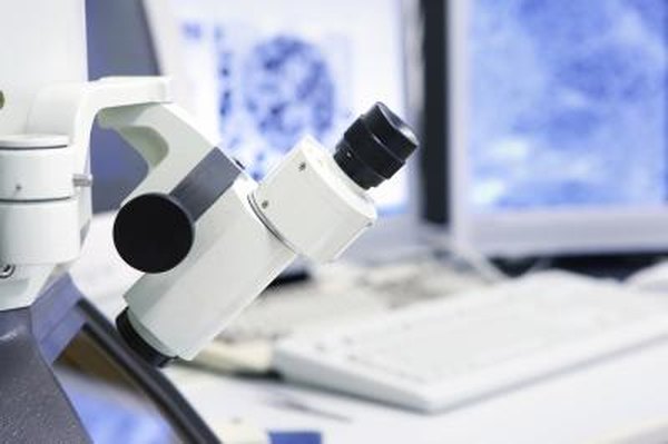
Dissecting microscope diagram with labels. Everything You Need to Know About A Dissecting Microscope A dissecting microscope focuses solely on the outer surface of the specimen, mostly of opaque objects that light can't pass through. It also does not offer high magnification unlike any other microscope. Its function is to give the viewer a wider field of vision all the while being able to work with it in real time. Label Microscope Diagram - EnchantedLearning.com arm - this attaches the eyepiece and body tube to the base. base - this supports the microscope. body tube - the tube that supports the eyepiece. coarse focus adjustment - a knob that makes large adjustments to the focus. diaphragm - an adjustable opening under the stage, allowing different amounts of light onto the stage. eyepiece - where you place your eye. Label the microscope — Science Learning Hub Use this interactive to identify and label the main parts of a microscope. Drag and drop the text labels onto the microscope diagram. eye piece lens: The lens you look through - normally 10x or 15x magnification. eye piece lens. coarse focus adjustment: Moves the lens up or down and adjusts focus. coarse focus adjustment. Parts of Dissecting Microscope | Botany - Biology Discussion In this article we will discuss about the parts of dissecting microscope with its working and utility. 1. Foot or Base: ADVERTISEMENTS: It is the basal, horse-shoe shaped or circular part of dissecting microscope. It is made of heavy material. It provides support to other parts of microscope.
› pmc › articlesAdvanced Fluorescence Microscopy Techniques—FRAP, FLIP, FLAP ... Apr 02, 2012 · 1. Introduction. FRAP, FLIP, FLAP, FRET, and FLIM are fluorescence microscopy techniques that in some way take advantage of particular aspects of the fluorescence process by which fluorochromes are excited and emit fluorescent light, are damaged during repetitive excitation, or undergo non-radiative decay prior to light emission. Dissecting microscope (Stereoscopic or Stereo microscope) Labeled Diagram of Dissecting microscope (Stereoscopic and Stereo microscope) A typical stereo microscope has 6 major parts which are:. LED Illuminators: Typically dissecting microscopes have an LED light that that illuminates the exhibit that needs to be observed. Eyepiece: Each dissecting microscope has two eyepieces that is used to focus on the light has divergent pathways. Dissection Microscopes The dissecting microscope, also known as a stereo microscope, might look like your run of the mill compound microscope but there are some very important differences between the two. While a dissecting microscope has a binocular eyepiece like a compound microscope, the image you get from a stereo microscope is, like its name implies ... Microscope Parts, Function, & Labeled Diagram - slidingmotion Objective lenses. Objective lenses are the most important part of the microscope. Its purpose is to visualize the specimen. There are 3-4 types of different objective lenses in any microscope. It has a magnification power of 4X to 100 X. 4X objective lens is the shortest lens while the 100X lens is the longest in terms of visualization.
rsscience.com › stereo-microscopeParts of Stereo Microscope (Dissecting microscope) - Rs' Science Stereo microscopes (also called Dissecting microscope) are branched out from other light microscopes for the application of viewing "3D" objects. These include substantial specimens, such as insects, feathers, leaves, rocks, sand grains, gems, coins, and stamps, etc. Functionally, a stereo microscope is like a powerful magnifying glass. 16 Parts of a Compound Microscope: Diagrams and Video Once you have an understanding of the parts of the microscope it will be much easier to navigate around and begin observing your specimen, which is the fun part! The 16 core parts of a compound microscope are: Head (Body) Arm. Base. Eyepiece. Eyepiece tube. dissecting microscope diagram - sqlinsaneo.com Base 16. Auxiliary lenses can be added to the objective lens to modify the magnification at the camera and the binocular eyetubes. Answer the following questions for each of your Microscope Label Diagram - 34 label diagram of microscope labels ... Microscope Label Diagram - 18 images - unbiology6, biology 521 resources, label a microscope teaching resources, quia protist vocabulary, Menu ≡ ╳ Home
30 Label The Parts And Functions Of The Microscope - Labels Database 2020
Dissecting Stereo Microscope Parts and Functions Other parts of a dissecting microscope: The head/body of a dissecting microscope contains several important components that are hidden within the tube. These include: · Prism - bend light and thus change the orientation of the image. · Relay lens - serve to invert the image and also extend the imaging system.
Post a Comment for "39 dissecting microscope diagram with labels"