42 heart diagram with labels and blood flow
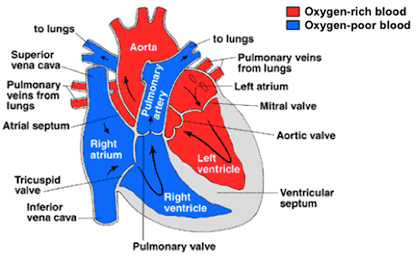
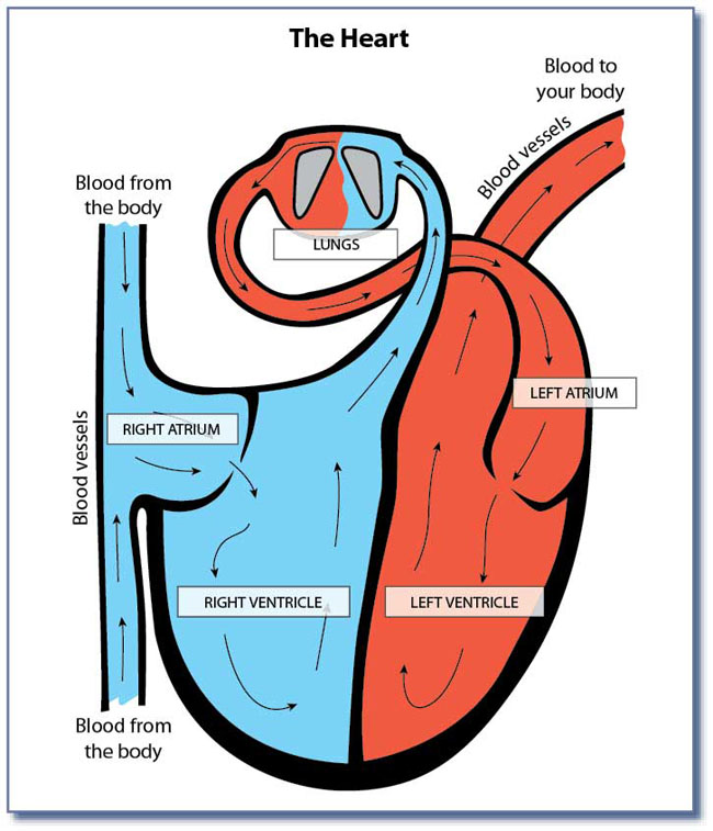
A Diagram of the Heart and Its Functioning Explained in Detail The heart blood flow diagram (flowchart) given below will help you to understand the pathway of blood through the heart.Initial five points denotes impure or deoxygenated blood and the last five points denotes pure or oxygenated blood. 1.Different Parts of the Body ↓ 2.Major Veins ↓ 3.Right Atrium ↓ 4.Right Ventricle ↓ 5.Pulmonary Artery ↓ 6.Lungs Circulatory System Diagram - New Health Advisor A systemic circuit represents the flow of blood around the organs and then movement of deoxygenated blood (which contains carbon dioxide and other waste materials) back to the heart. ... There are different types of circulatory system diagrams; some have labels while others don't. The color blue stands for deoxygenated blood while red stands ...
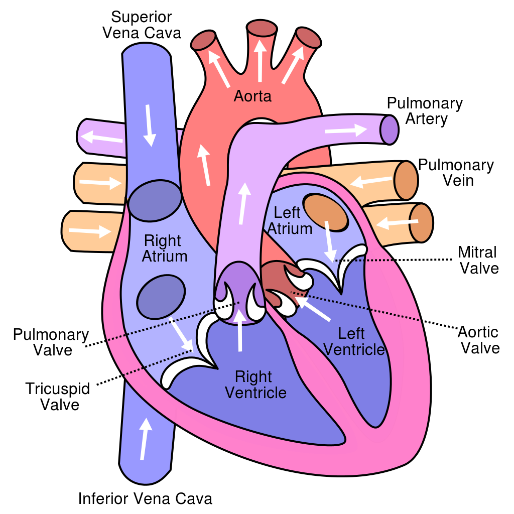
Heart Diagram with Labels and Detailed Explanation Heart Detailed Diagram Heart - Chambers There are four chambers of the heart . The upper two chambers are the auricles and the lower two are called ventricles. The two atria are thin-walled chambers that receive blood from the veins. The two ventricles are in contrast thick-walled which forcefully pump blood out of the heart.
Heart diagram with labels and blood flow
HEART DIAGRAM Flashcards | Quizlet Now up your study game with Learn mode. Try Learn mode. Study with Flashcards again. 1/28. Created by. derekhl62. Labels of the Heart and Blood Flow Info. Upgrade to remove ads. Only $2.99/month. › 1-label-the-heartLabel the heart — Science Learning Hub Jun 16, 2017 · Labels. Description. Vena cava. Carries deoxygenated blood from the body to the heart. Semilunar valve. Flaps that prevent backflow of blood. Left atrium. Receives oxygenated blood from the lungs. Left ventricle. Region of the heart that pumps oxygenated blood to the body. Pulmonary artery. Carries deoxygenated blood to the lungs. Right ventricle Blood Flow Through The Body Diagram stock illustrations Vector isolated illustration of human internal organs and circulatory system in man body. Stomach, liver, bladder, lung, kidney, heart, icon. Medical poster. The human cardiovascular system (also known as the circulatory system). A medical diagram showing the heart, arteries and veins of the human body. heart anatomy.
Heart diagram with labels and blood flow. Heart Anatomy: Labeled Diagram, Structures, Blood Flow ... - EZmed There are 4 chambers, labeled 1-4 on the diagram below. To help simplify things, we can convert the heart into a square. We will then divide that square into 4 different boxes which will represent the 4 chambers of the heart. The boxes are numbered to correlate with the labeled chambers on the cartoon diagram. › photos › muscular-systemMuscular System Labeled Diagram Stock Photos, Pictures ... Heart blood flow circulation anatomical diagram with atrium and ventricle system. Vector illustration labeled medical poster. Heart blood flow anatomical diagram with atrium and ventricle system. Vector illustration labeled medical poster. Blood circulation path scheme with arrows. muscular system labeled diagram stock illustrations Heart Anatomy & Circulatory System Blood Flow Two labeled diagrams presenting the anatomy of the heart and the circulatory systems. The top right provides an in-depth diagram of the anatomy of the chambers, valves, and vasculature connected to the heart. The bottom left emphasizes the anatomy of the systemic and pulmonary circulations. lsa.colorado.edu › essence › textsThe Heart and Circulation of Blood - LSA The center of the circulatory system is the heart, which is the main pumping mechanism. The heart is made of muscle. The heart is shaped something like a cone, with a pointed bottom and a round top. It is hollow so that it can fill up with blood. An adult’s heart is about the size of a large orange and weighs a little less than a pound.
Box Diagram, Labels of Heart, and Blood Flow through Heart About Press Copyright Contact us Creators Advertise Developers Terms Privacy Policy & Safety How YouTube works Test new features Press Copyright Contact us Creators ... File:Heart diagram blood flow en.svg - Wikipedia File:Heart diagram blood flow en.svg. Size of this PNG preview of this SVG file: 330 × 370 pixels. Other resolutions: 214 × 240 pixels | 428 × 480 pixels | 535 × 600 pixels | 685 × 768 pixels | 913 × 1,024 pixels | 1,827 × 2,048 pixels. This is a file from the Wikimedia Commons. Information from its description page there is shown below. Heart Diagram Flow Teaching Resources | Teachers Pay Teachers Cardiovascular System: Heart Diagram to Color by Lori Maldonado 80 $2.00 PDF This diagram shows the way blood flows through the heart. The areas of the heart with MORE oxygen are labeled with an "R". Students will color these areas RED. The areas of the heart with LESS oxygen are labeled with a "B". Students will color these areas BLUE. › heart › picture-of-the-heartHuman Heart (Anatomy): Diagram, Function, Chambers, Location ... This lets blood flow better and can abort a heart attack or relieve angina (chest pain). Thrombolysis : “Clot-busting” drugs injected into the veins can dissolve a blood clot causing a heart ...
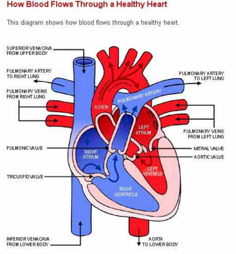
Diagram of Blood Flow in the Heart, Lungs and Body This medical illustration depicts a diagram of blood flow through the body. Oxygenated (oxygen-rich)blood travels from the lungs to the heart, where it is then pumped throughout the body. Deoxygenated (oxygen-poor) blood travels from the body back to the heart, where it is pumped to the lungs for gas exchange. Labels include the common carotid arteries, jugular veins, superior vena cava ... › pmc › articlesCerebral blood flow and autoregulation: current measurement ... Jun 21, 2016 · 1.1. Physiological importance and normal values of cerebral blood flow in adult humans. The human brain is an organ with high-energy density demands, amounting to only 2% of the entire body mass (or ∼ 1.4 kg) but accounting for about 20% of the total power consumption of a normal adult at rest (or ∼ 20 W). PDF BLOOD FLOW THROUGH THE HEART diagram deoxygenated blood from body tissue oxygenated blood from lungs via pulmonary vein s superior and inferior vena cava left atrium right atrium bicuspid valve tricuspid valve left ventricle ... blood flow through the heart diagram author: taustin created date: 9/30/2013 9:09:13 pm byjus.com › biology › human-heartHuman Heart - Anatomy, Functions and Facts about Heart The external structure of the heart has many blood vessels that form a network, with other major vessels emerging from within the structure. The blood vessels typically comprise the following: Veins supply deoxygenated blood to the heart via inferior and superior vena cava, and it eventually drains into the right atrium.
Heart Arrhythmias: An Exercise Professional’s Primer
Heart Blood Flow | Simple Anatomy Diagram, Cardiac Circulation Pathway ... Step 1 and 6 involve a blood vessel, which makes sense as this is how blood enters and exits that side of the heart. Steps 2-5 involve a chamber, valve, chamber, and valve. So if you remember this general pattern, it will help you recall the order in which blood flows through each side of the heart. Right Side of the Heart SVC/IVC Right Atrium
Tips for How to Study the Cardiovascular System
PDF Heart Diagram Blood Flow Quiz Free Books [READ] Heart Diagram Blood Flow Quiz PDF Book is the book you are looking for, by download PDF Heart Diagram Blood Flow Quiz book you are also motivated to ... Label Blood Flow Through The Heart Quiz Label Blood Flow Through The Heart Quiz Heart Health Types Causes Symptoms Amp Treatments, Di Feb 12th, 2022
How the Heart Works | Congenital Heart Defects | NCBDDD | CDC
Blood Circulation In Heart Flowchart in 14 Steps Blood circulation in the heart flowchart is divided into the left and right sides. The right side is the flow of deoxygenated blood. The left side represents the flow of oxygenated blood. There are 14 steps in a circle of blood for easy illustration: Deoxygenated blood starts to run from the body It flows into Superior/ Inferior Vena Cava

Heart Anatomy Blood Flow - Anatomy Drawing Diagram
Heart Diagram with Labels and Detailed Explanation - BYJUS The diagram of heart is beneficial for Class 10 and 12 and is frequently asked in the examinations. A detailed explanation of the heart along with a well-labelled diagram is given for reference. Well-Labelled Diagram of Heart The heart is made up of four chambers: The upper two chambers of the heart are called auricles.
Circulatory System Diagram | New Health Advisor
Circulatory System: Blood Flow Pathway Through the Heart Pathway of Blood Through the Heart. In this educational lesson, we learn about the blood flow order through the human heart in 14 easy steps, from the superior and inferior vena cava to the atria and ventricles. Come also learn with us the heart's anatomy, including where deoxygenated and oxygenated blood flow, in the superior vena cava, inferior vena cava, atrium, ventricle, aorta ...
Heart Diagram with Blood Flow - peshsexam2
Heart Diagrams for Labeling and Coloring, With ... - Teachers Pay Teachers - One black and white heart diagram with lines for students to fill in labels, and arrows showing blood flow - One black and white heart diagram with no lines or labels, but arrows included, so you can customize what labels the diagram will include - One black and white heart diagram with no lines, labels, or arrows, but with texture of the ...
Mike Tyson Tattoos: Heart Diagram Blood Flow
The Heart - Science Quiz - GeoGuessr This science quiz game will help you identify the parts of the human heart with ease. Blood comes in through veins and exists via arteries—to control the direction of the flow, the heart has four sets of valves. The heart is an amazing machine with a lot of moving parts—let this quiz game help you find your way around this most vital of organs.

Pin on EMS, EMT and Paramedics
Circulatory System Diagram - Cardiovascular System and Blood ... SmartDraw has a number of templates included for circulatory system diagrams, cardiovascular system diagrams, blood circulation diagrams, and more. You don't really have to "draw" them as much as find them and modify them as needed. You can add labels or titles and change the size of symbols as necessary.

Alila Medical Media | Heart and Circulatory System Images
Diagram of heart---Pathway flow of blood - Liveworksheets Diagram of heart---Pathway flow of blood label the heart diagram ID: 1791581 Language: English School subject: health Grade/level: 9-10 Age: 14-15 Main content: Heart diagram Other contents: Add to my workbooks (2) Download file pdf Embed in my website or blog Add to Google Classroom

Brenda's A & P Eportfolio: Objective 55: Trace the path of blood flow through the kidneys
The Heart | Circulatory Anatomy - Visible Body One chamber on the left receives oxygen-rich blood from the lungs and another pumps that nutrient-rich blood into the body. Two valves control blood flow within the heart's chambers, and two valves control blood flow out of the heart. 1. The Heart Wall Is Composed of Three Layers. The muscular wall of the heart has three layers.

Adult Heart Diagram - Milf Nude Photo
Human Heart Diagram Labeled | Science Trends Let's examine the anatomy of the heart along with some diagrams that show how the heart operates. Anatomy Of The Heart The human heart usually weighs somewhere between 10 to 12 ounces in men and between 8 to 10 ounces in women, and in terms of size is roughly the size of the fist.
Heart Diagram With Labels And Blood Flow | MedicineBTG.com
Diagram of Blood Flow Through the Heart - Bodytomy The heart is divided into two chambers, left and right, the right atrium and ventricle lie on the right side and the left atrium and ventricle on the left side. These two chambers are not directly connected to each other. Synchronization of the Two Chamber The right and left side or chambers of the heart work in tandem with each other.

Blood Flow Through The Heart Stock Photo: 7710767 - Alamy
labeled diagram of circulatory system Diagram of Heart Blood Flow for Cardiac Nursing Students - NCLEX Quiz we have 12 Pics about Diagram of Heart Blood Flow for Cardiac Nursing Students - NCLEX Quiz like Performance Vein Institute of Los Alamitos: Chronic Venous, The Anatomy Collection - Palace Learning and also Circulatory System Diagram | Anatomy and physiology, Human circulatory.

PPT - The Heart PowerPoint Presentation - ID:2746716
pmt.physicsandmathstutor.com › download › BiologyPractical notes - SP 2.3c Dissection of a Mammalian Heart ... 3. Use a glass rod to follow the path of blood flow : via the pulmonary vein, left atrium and through the bicuspid valve into the left ventricle; via the left ventricle through the semilunar valve and out of the aorta. 4. Note the muscular surface of the ventricle chambers which ensur es smooth blood flow. 5.

Heart Anatomy Blood Flow - Anatomy Drawing Diagram
Heart-diagram-with-labels-heart-diagram-blood-flow ... - Course Hero View heart-diagram-with-labels-heart-diagram-blood-flow-originalstylophone-of-heart-diagram-with-labels.j from BIO 2070 at College of Southern Maryland. From body Aorta To lung Superior vena
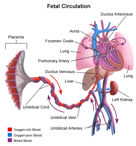
Fetal Circulation
Diagram of Human Heart and Blood Circulation in It Every heart diagram labeledwill clearly show these valves. These valves allow blood flow in one direction only. Different valves perform different functions. Tricuspid valve is located between the right ventricle of your heart and the right atrium, and allows the blood to move from the right atrium to the right ventricle.
Post a Comment for "42 heart diagram with labels and blood flow"