44 cell structure with labels
Bacteria in Microbiology - shapes, structure and diagram The bacteria shapes, structure, and labeled diagrams are discussed below. Sizes The sizes of bacteria cells that can infect human beings range from 0.1 to 10 micrometers. Some larger types of bacteria such as the rickettsias, mycoplasmas, and chlamydias have similar sizes as the largest types of viruses, the poxviruses. Cell: Structure and Functions (With Diagram) - Biology Discussion Eukaryotic Cells: 1. Eukaryotes are sophisticated cells with a well defined nucleus and cell organelles. 2. The cells are comparatively larger in size (10-100 μm). 3. Unicellular to multicellular in nature and evolved ~1 billion years ago. 4. The cell membrane is semipermeable and flexible. 5. These cells reproduce both asexually and sexually.
A Labeled Diagram of the Animal Cell and its Organelles A Labeled Diagram of the Animal Cell and its Organelles There are two types of cells - Prokaryotic and Eucaryotic. Eukaryotic cells are larger, more complex, and have evolved more recently than prokaryotes. Where, prokaryotes are just bacteria and archaea, eukaryotes are literally everything else.

Cell structure with labels
Human Cell Diagram, Parts, Pictures, Structure and Functions Human Cell Diagram, Parts, Pictures, Structure and Functions. The cell is the basic functional in a human meaning that it is a self-contained and fully operational living entity. Humans are multicellular organisms with various different types of cells that work together to sustain life. Other non-cellular components in the body include water ... Microscope Imaging Station. Gallery. - Exploratorium Some plant cells have organelles called chloroplasts that make them green and able to capture energy from light. Rigid walls typically made of cellulose surround plant cells. Video: Elodea leaf cells with structures labeled Chloroplasts and mitochondria move within Elodea leaf cells; nuclei are also visible as clear, fried-egg-shaped structures. Plant Cell - Definition, Structure, Function, Diagram & Types It is a rigid layer which is composed of polysaccharides cellulose, pectin and hemicellulose. It is located outside the cell membrane. It also comprises glycoproteins and polymers such as lignin, cutin, or suberin. The primary function of the cell wall is to protect and provide structural support to the cell.
Cell structure with labels. Labeled Plant Cell With Diagrams | Science Trends The parts of a plant cell include the cell wall, the cell membrane, the cytoskeleton or cytoplasm, the nucleus, the Golgi body, the mitochondria, the peroxisome's, the vacuoles, ribosomes, and the endoplasmic reticulum. Parts Of A Plant Cell The Cell Wall Let's start from the outside and work our way inwards. Learn the parts of a cell with diagrams and cell quizzes - Kenhub Two major regions can be found in a cell. The first is the cell nucleus, which houses DNA in the form of chromosomes. The second is the cytoplasm, a thick solution mainly comprised of water, salts, and proteins. The parts of a eukaryotic cell responsible for maintaining cell homeostasis, known as organelles, are located within the cytoplasm. Animal Cells: Labelled Diagram, Definitions, and Structure The endoplasmic reticulum (s) are organelles that create a network of membranes that transport substances around the cell. They have phospholipid bilayers. There are two types of ER: the rough ER, and the smooth ER. The rough endoplasmic reticulum is rough because it has ribosomes (which is explained below) attached to it. Cell Structure | Thermo Fisher Scientific - US Cell Structure. Organelle structures play a critical role in cellular function, and the detection of specific cell structures is key in fluorescence imaging of cells and tissue. Cell structure labels can be a counterstain method to identify the location of specific proteins and targets of interest within the cell.
Cell Organelles- Definition, Structure, Functions, Diagram In a plant cell, the cell wall is made up of cellulose, hemicellulose, and proteins while in a fungal cell, it is composed of chitin. A cell wall is multilayered with a middle lamina, a primary cell wall, and a secondary cell wall. The middle lamina contains polysaccharides that provide adhesion and allow binding of the cells to one another. Bacteria Cell Structures with Labels Stock Vector - Dreamstime Bacteria Cell Structures with labels Royalty-Free Vector Bacterial cell structures labeled on a bacillus cell with nucleoid DNA and ribosomes. External structures include the capsule, pili, and flagellum. Morphology of internal structures of bacteria. cell anatomy bacteria, prokaryotic cell, cell, internal structures, prokaryotic, dna, bacteria, Bacterial Cell Structure Labeling Diagram | Quizlet Cell Membrane A thin sheet of lipid and protein that surrounds the cytoplasm and controls the flow of materials in and out of the cell S Layer Monolayer of protein used for protection and attachment Fimbrae Fine bristles extending from cell surface that help in adhesion to other cells and surfaces. Outer Membrane Cell - Label | Cell Structure Quiz - Quizizz 16 Questions Show answers Question 1 30 seconds Q. Label #3 answer choices Nucleus Mitochondria Vacuole Golgi Body Question 2 30 seconds Q. Label #4 answer choices Cell wall Cell membrane Nuclear membrane Gatekeeper Question 3 30 seconds Q. Label #5 answer choices Nucleus Nucleolus Endoplasmic Reticulum Mitochondria Question 4 30 seconds
Cell Structure | SEER Training The nucleus determines how the cell will function, as well as the basic structure of that cell. Cytoplasm. The cytoplasm is the gel-like fluid inside the cell. It is the medium for chemical reaction. It provides a platform upon which other organelles can operate within the cell. All of the functions for cell expansion, growth and replication ... Label Cell Parts | Plant & Animal Cell Activity | StoryboardThat Click "Start Assignment". Find diagrams of a plant and an animal cell in the Science tab. Using arrows and Textables, label each part of the cell and describe its function. Color the text boxes to group them into organelles found in only animal cells, organelles found in only plant cells, and organelles found in both cell types. Plant Cells: Labelled Diagram, Definitions, and Structure The cell wall is made of cellulose and lignin, which are strong and tough compounds. Plant Cells Labelled Plastids and Chloroplasts Plants make their own food through photosynthesis. Plant cells have plastids, which animal cells don't. Plastids are organelles used to make and store needed compounds. Chloroplasts are the most important of plastids. File:Plant cell structure svg labels.svg - Wikipedia File:Plant cell structure svg labels.svg. Size of this PNG preview of this SVG file: 649 × 475 pixels. Other resolutions: 320 × 234 pixels | 640 × 468 pixels | 1,024 × 749 pixels | 1,280 × 937 pixels | 2,560 × 1,874 pixels. This is a file from the Wikimedia Commons. Information from its description page there is shown below.
Student Cell Model | Matthew | Flickr
Plant Cell- Definition, Structure, Parts, Functions, Labeled Diagram The central vacuoles are found in the cytoplasmic layer of cells of a variety of different organisms, but larger in the plant cells. Structure of plant cell vacuoles. These are large, vesicles filled with fluid, within the cytoplasm of a cell. It is made up of 30% fluid of the cell volume but can fill up to 90% of the cell's intracellular space.
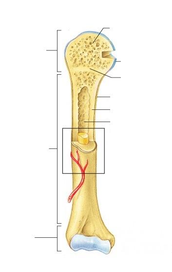
Exercise 9: Overview of the Skeleton: Classification and Structure of ...
Structure of Bacterial Cell (With Diagram) - Biology Discussion It is 10-25 nm in thickness. It gives shape to the cell. Nucleus: The single circular double-stranded chromosome is the bacterial genome. Other structures include cytoplasmic membrane, mesosomes, ribosomes and cytoplasmic inclusions. Unlike eukaryotes cytoplasm does not contain ribosome, Golgi, cytoskeleton.
SQL Workbench/J User's Manual SQLWorkbench
Cathodoluminescence imaging of cellular structures labeled with ... However, in cells that were pre-fixed before IRAZOLVE-MITO application, the staining pattern changed and the granular and filamentous structures extending throughout the cells were labeled (Fig. 1).

Exploration of the Human Spinal Cord
Plant and Animal Cell: Labeled Diagram, Structure, Function - Embibe Double membrane-bound structures found only in the plant cells. 2. This is an autonomous organelle. 3. There are stroma or matrix and grana or stacked discs that are involved in photosynthesis. 4. Grana are the site for photochemical reactions of photosynthesis, while stroma is the site for biochemical reactions of photosynthesis.

Eukaryotic Cell Labeling - PurposeGames
A Labeled Diagram of the Plant Cell and Functions of its Organelles A Labeled Plant Cell Amyloplasts A major component of plants that are starchy in nature, the amyloplasts are organelles that store starch. They are classified as plastids, and are also known as starch grains. They are responsible for the conversion of starch into sugar, that gives energy to the starchy plants and tubers.

Bismuth trisulfide (Bi2S3) | PVEducation
Animal Cell Diagram with Label and Explanation: Cell Structure, Functions Animal cell is a typical Eukaryotic cell enclosed by a plasma membrane containing nucleus and organelles which lack cell walls, unlike all other Eukaryotic cells. The typical cell ranges in size between 1-100 micrometers. The lack of cell walls enabled the animal cells to develop a greater diversity of cell types.
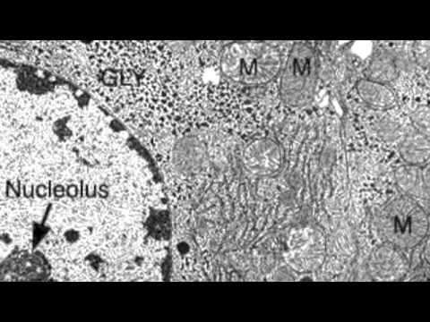
2.3.3 Identify structures from electron micrographs of liver cells ...
Label the cell structure. | Study.com Label the cell structure. Cells: All living cells contain an intracellular space called the cytoplasm. The cytoplasm is filled with a jelly-like fluid where many of the cells enzymatic reactions...

Animal Cells Diagram with Labels Awesome Animal Cell Diagrams Labeled ...
Cell Organelles - Types, Structure and their Functions Ribosomes are found in the form of tiny particles in a large number of cells and are mainly composed of 2/3rd of RNA and 1/3rd of protein. They are named as the 70s (found in prokaryotes) or 80s (found in eukaryotes) The letter S refers to the density and the size, known as Svedberg's Unit. Both 70S and 80S ribosomes are composed of two subunits.
Post a Comment for "44 cell structure with labels"